- Department of Neurosurgery, Kagoshima University, Kagoshima, Japan
- Department of Neurosurgery, Izumi Regional Hospital, Akune, Kagoshima, Japan
- Department of Clinical Radiology, Izumi Regional Hospital, Akune, Kagoshima, Japan
- Department of Neurosurgery, Mayo Clinic Hospital, Rochester, United States
Correspondence Address:
Kazunori Arita, Department of Neurosurgery, Kagoshima University, Kagoshima City, Kagoshima Prefecture, Japan.
DOI:10.25259/SNI_559_2024
Copyright: © 2024 Surgical Neurology International This is an open-access article distributed under the terms of the Creative Commons Attribution-Non Commercial-Share Alike 4.0 License, which allows others to remix, transform, and build upon the work non-commercially, as long as the author is credited and the new creations are licensed under the identical terms.How to cite this article: Eri Inoue1,2, Shingo Fujio1, Hiroshi Hosoyama1,2, Shinichiro Yoshimura3, FM Moinuddin4, Ryosuke Hanaya1, Kazunori Arita1,2. A pituitary gland squeezed upward by intrasellar kissing carotid arteries: Mimicking a pituitary microadenoma. 11-Oct-2024;15:372
How to cite this URL: Eri Inoue1,2, Shingo Fujio1, Hiroshi Hosoyama1,2, Shinichiro Yoshimura3, FM Moinuddin4, Ryosuke Hanaya1, Kazunori Arita1,2. A pituitary gland squeezed upward by intrasellar kissing carotid arteries: Mimicking a pituitary microadenoma. 11-Oct-2024;15:372. Available from: https://surgicalneurologyint.com/?post_type=surgicalint_articles&p=13143
Date of Submission
08-Jul-2024
Date of Acceptance
14-Sep-2024
Date of Web Publication
11-Oct-2024
Abstract
Background: Intrasellar kissing carotid arteries are a rare variant in which bilateral internal carotid arteries run very near each other at their cavernous sinus portion. We encountered a woman with the pituitary gland mimicking a pituitary microadenoma because the pituitary gland was compressed bilaterally by intrasellar kissing carotid arteries.
Case Description: A 61-year-old woman with a chronic headache underwent magnetic resonance imaging, which revealed a sellar mass measuring 10.2 mm in height, 8.2 mm in length, and 4.0 mm in width at the midintercarotid level. Blood levels of all pituitary and target-organ hormones were within normal range. The height and superior convex shape of the sellar mass suggested that it was a nonfunctioning microadenoma, which was monitored over the past 16 years. A recent three-dimensional reconstruction of magnetic resonance angiography clearly showed that the pituitary gland was squeezed upward, compressed bilaterally, and extended superiorly by intrasellar kissing carotid arteries.
Conclusion: The pituitary gland can be squeezed upward by intrasellar kissing carotid arteries and mimic pituitary tumor.
Keywords: Intrasellar kissing carotid arteries, Pituitary gland, Pituitary microadenoma, Pituitary tumor
INTRODUCTION
In general, the size of human pituitary glands typically increases during childhood and adolescence, reaching its peak in the second or third decade of life.[
We hereby report a postmenopausal woman with a pituitary gland of 10.2 mm in height with a convex superior surface, which was accompanied by intrasellar kissing carotid arteries. The case has been followed for 16 years under the suspicion of micro-non-functioning pituitary adenoma.
CASE DESCRIPTION
A 61-year-old woman with a chronic headache underwent magnetic resonance imaging (MRI), which suggested the presence of a sellar mass. The mass showed an intensity similar to that of cerebral gray matter on both T1-weighted (T1W) and T2-weighted MRI [

Figure 1:
Magnetic resonance imaging of the pituitary mass at 61 years old. (a) T1-weighted (W) sagittal image. (b) T1W axial image. (c) T2W coronal image. (d) Gadolinium-enhanced (GE) sagittal image. (e) GE axial image. (f) GE coronal image. Red bar in 1a: Antero-posterior diameter, Yellow bar in 1d: Height, Blue bar: Transverse diameter at mid-intercarotid level. A pituitary mass (arrow, in a-c) with similar intensity to the brain parenchyma existed in front of the posterior pituitary lobe (arrowhead in a and b). The gadolinium moderately and diffusely enhanced the pituitary mass (arrow, in d-f).
Young neurosurgeons initially in charge of this patient suggested the mass to be the pituitary gland harboring nonfunctioning pituitary microadenoma due to the height and convex upper surface.
The patient was initially followed up annually and later every few years. However, recent MRI studies using 3T-machine (SIGNA, GE Healthcare, US) revealed that the mass remained unchanged over the 16 years [
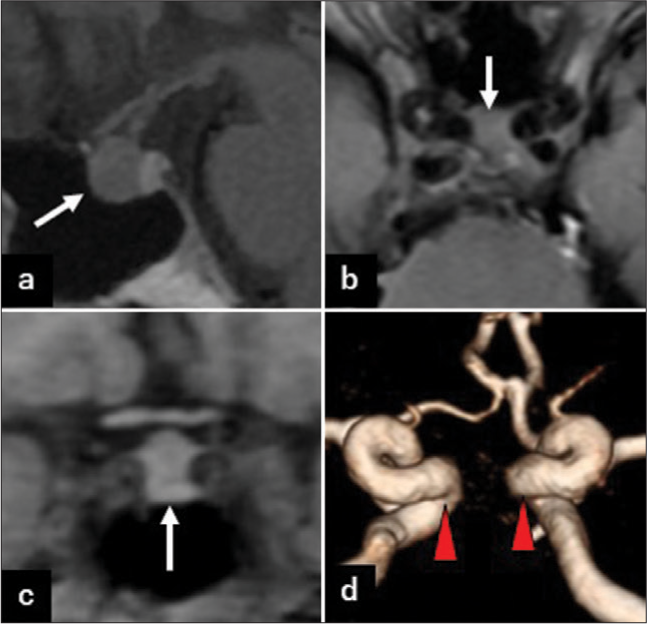
Figure 2:
Magnetic resonance imaging of the pituitary mass at 77 years old. (a) T1-weighted (W) sagittal image. (b) T1W axial image. (c) T1W coronal image. (d) Three-dimensional reconstruction of time-of-flight magnetic resonance angiography. The size of the pituitary mass (arrow) did not change compared to 16 years before (a-c). Bilateral tortuous internal carotid arteries (red arrowheads) are running very near to each other (d).
Three-dimensional (3D) reconstruction of magnetic resonance angiography showed that the kissing carotid arteries at the sella turcica were squeezing the pituitary gland [

Figure 3:
Magnetic resonance imaging-based three-dimensional reconstruction images of the pituitary gland and bilateral internal carotid arteries (IC). (a) Anteroposterior view, (b) left anterior oblique view, (c) left-right view, (d) right anterior oblique view, (e) right-left view, and (f) posteroanterior view. Blue: Anterior pituitary gland, Pink: Pituitary stalk, PL: Posterior pituitary lobe, Bar: 10 mm. Anterior pituitary gland is compressed by the “intrasellar kissing carotid arteries” and squeezed upward.
DISCUSSION
In this case, the height of the pituitary gland was 10.2 mm at the time of initial MRI.
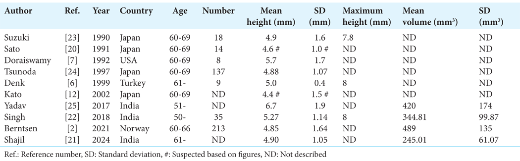
On the other hand, the average volume of the pituitary gland in females older than 50 is reported to range from 245.1 to 489 mm3 [
How to explain the discrepancy between the abnormal height and convex pituitary surface and the normal volume of the pituitary gland? Reported mean intercarotid distances at the sellar region varied considerably, from 11.4 to 17.1 mm, due to the differences in research subjects, which include cadavers, autopsy specimens, and neuroimaging of healthy adults, as well as variations in measuring methods [
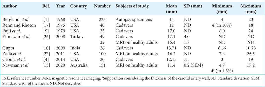
However, the distance can sometimes become very narrow, 4 mm or less, due to the tortuous course of the internal carotid arteries, intrasellar kissing carotid arteries. In Renn and Rhoton’s cadaveric study, the narrowest distance was 4 mm in 10% of 40 cadavers.[
In this case, a recent follow-up MRI clearly showed that the pituitary gland was bilaterally compressed by intrasellar kissing carotid arteries. Perhaps a large opening of diaphragma sellae, as observed in 40% of cadaveric specimens,[
CONCLUSION
The authors presented an elderly woman with intrasellar kissing carotid arteries who was suspected of having a pituitary microadenoma based on the height and shape of the pituitary gland on MRI. However, over 16 years, the lesion has not grown. We finally concluded that this is the normal pituitary gland, which is squeezed upward by the kissing carotid arteries.
Ethical approval
The Institutional Review Board has waived the ethical approval for this study.
Declaration of patient consent
The authors certify that they have obtained all appropriate patient consent.
Financial support and sponsorship
Nil.
Conflicts of interest
There are no conflicts of interest.
Use of artificial intelligence (AI)-assisted technology for manuscript preparation
The authors confirm that there was no use of artificial intelligence (AI)-assisted technology for assisting in the writing or editing of the manuscript and no images were manipulated using AI.
Disclaimer
The views and opinions expressed in this article are those of the authors and do not necessarily reflect the official policy or position of the Journal or its management. The information contained in this article should not be considered to be medical advice; patients should consult their own physicians for advice as to their specific medical needs.
References
1. Bergland RM, Ray BS, Torack RM. Anatomical variations in the pituitary gland and adjacent structures in 225 human autopsy cases. J Neurosurg. 1968. 28: 93-9
2. Berntsen EM, Haukedal MD, Håberg AK. Normative data for pituitary size and volume in the general population between 50 and 66 years. Pituitary. 2021. 24: 737-45
3. Campero A, Martins C, Yasuda A, Rhoton AL. Microsurgical anatomy of the diaphragma sellae and its role in directing the pattern of growth of pituitary adenomas. Neurosurgery. 2008. 62: 717-23
4. Cebula H, Kurbanov A, Zimmer LA, Poczos P, Leach JL, De Battista JC. Endoscopic, endonasal variability in the anatomy of the internal carotid artery. World Neurosurg. 2014. 82: e759-64
5. Chanson P, Daujat F, Young J, Bellucci A, Kujas M, Doyon D. Normal pituitary hypertrophy as a frequent cause of pituitary incidentaloma: A follow-up study. J Clin Endocrinol Metab. 2001. 86: 3009-15
6. Denk CC, Onderoğlu S, Ilgi S, Gürcan F. Height of normal pituitary gland on MRI: Differences between age groups and sexes. Okajimas Folia Anat Jpn. 1999. 76: 81-7
7. Doraiswamy PM, Potts JM, Axelson DA, Husain MM, Lurie SN, Na C. MR assessment of pituitary gland morphology in healthy volunteers: Age-and gender-related differences. AJNR Am J Neuroradiol. 1992. 13: 1295-9
8. Elster AD, Chen MY, Williams DW, Key LL. Pituitary gland: MR imaging of physiologic hypertrophy in adolescence. Radiology. 1990. 174: 681-5
9. Fujii K, Chambers SM, Rhoton AL. Neurovascular relationships of the sphenoid sinus. A microsurgical study. J Neurosurg. 1979. 50: 31-9
10. Gupta T. An anatomical study of intercarotid distances in the sellar region with a surgical perspective. Braz J Morphol Sci. 2009. 26: 23-6
11. Ikram MF, Sajjad Z, Shokh I, Omair A. Pituitary height on magnetic resonance imaging observation of age and sex related changes. J Pak Med Assoc. 2008. 58: 261-5
12. Kato K, Saeki N, Yamaura A. Morphological changes on MR imaging of the normal pituitary gland related to age and sex: main emphasis on pubescent females. J Clin Neurosci. 2002. 9: 53-6
13. Laws ER, Kern EB. Complications of trans-sphenoidal surgery. Clin Neurosurg. 1976. 23: 401-16
14. Lurie SN, Doraiswamy PM, Husain MM, Boyko OB, Ellinwood EH, Figiel GS. In vivo assessment of pituitary gland volume with magnetic resonance imaging: the effect of age. J Clin Endocrinol Metab. 1990. 71: 505-8
15. Newman H, Milne N, Lewis SB. Neurosurgical anatomy of the internal carotid artery: Magnetic resonance imaging study of the Sellar region. World Neurosurg. 2020. 133: e711-5
16. Pereira Filho Ade A, Gobbato PL, Pereira Filho Gde A, Silva SB, Kraemer JL. Intracranial intrasellar kissing carotid arteries: Case report. Arq Neuropsiquiatr. 2007. 65: 355-7
17. Renn WH, Rhoton AL. Microsurgical anatomy of the sellar region. J Neurosurg. 1975. 43: 288-98
18. Saberi S, Bushman J, Sinha S, Shlensky D, Bapuraj J, Esfandiari NH. Kissing carotid arteries causing male hypogonadotropic hypogonadism. AACE Clin Case Rep. 2024. 10: 31-2
19. Sahin M, Dilli A, Karbek B, Unsal IO, Gungunes A, Colak N. Unusual cause of primary amenorrhea due to kissing internal carotid arteries. Pituitary. 2012. 15: 258-9
20. Sato Y. MRI of normal pituitary glands and their surrounding structures. J UOEH. 1991. 13: 295-311
21. Shajil S, Sharma PK, Sekar A, Rajendran G, Amir A. Role of magnetic resonance imaging in the evaluation of age-and gender-related changes in the dimensions of the pituitary gland in the Indian population. Cureus. 2024. 16: e54093
22. Singh AK, Kandasamy D, Garg A, Jyotsna VP, Khadgawat R. Study of pituitary morphometry using MRI in Indian subjects. Indian J Endocrinol Metab. 2018. 22: 605-9
23. Suzuki M, Takashima T, Kadoya M, Konishi H, Kameyama T, Yoshikawa J. Height of normal pituitary gland on MR imaging: Age and sex differentiation. J Comput Assist Tomogr. 1990. 14: 36-9
24. Tsunoda A, Okuda O, Sato K. MR height of the pituitary gland as a function of age and sex: Especially physiological hypertrophy in adolescence and in climacterium. AJNR Am J Neuroradiol. 1997. 18: 551-4
25. Yadav P, Singhal S, Chauhan S, Harit S. MRI Evaluation of size and shape of normal pituitary gland: Age and sex related changes. J Clin Diagn Res. 2017. 11: 1-4
26. Yilmazlar S, Kocaeli H, Eyigor O, Hakyemez B, Korfali E. Clinical importance of the basal cavernous sinuses and cavernous carotid arteries relative to the pituitary gland and macroadenomas: Quantitative analysis of the complete anatomy. Surg Neurol. 2008. 70: 165-74
27. Zada G, Agarwalla PK, Mukundan S, Dunn I, Golby AJ, Laws ER. The neurosurgical anatomy of the sphenoid sinus and sellar floor in endoscopic transsphenoidal surgery. J Neurosurg. 2011. 114: 1319-30